Nanomedicine: A Vast Horizon on a Molecular Landscape – Part VIII, Magnetic Nanoparticles theranostics
Mar 7th, 2017 by Jing Zhou | News | Recent News & Articles |
 This is the eighth article in a review series on Nanomedicine. We started from reviewing the major research and entrepreneurial development of nanomedicine and the relevant patent landscape (Part I and Part II). The first topic we discussed was Organs-on-a-chip (Part III). Following that, we focused on nanotechnology in medical therapeutics. Nanoparticles have nanoscale dimensions and demonstrate unique chemical and physical properties from their bulk. This also gives them great advantages in drug delivery (Part IV), cancer therapeutics (Part V), and bio-imaging (Part VI). In the last installment, we reviewed one special type of nanoparticles: quantum dots, which are incredibly small semiconductor particles (Part VII). Here, we will review the theranostic applications and IP landscape of another special type of nanoparticles known as magnetic nanoparticles (MNP). As in the past, those patent documents cited in the article are summarized in the table at the end.
Magnetic Nanoparticles
Magnetic nanoparticles, also known as superparamagnetic nanoparticles are small inorganic crystals about 5-20 nm in diameter. Two main classes of MNPs currently used for clinical imaging are ferromagnetic iron oxide nanoparticles and ultrasmall superparameganetic iron oxide nanoparticles (USPION). MNPs are usually multilayer materials, which give them their various properties and functionalities for diagnosis and disease treatment. The structure of iron oxide nanoparticles has three main components: an iron oxide core as a Magnetic Resonance Imaging (MRI) contrast agent, a biocompatible coating outside the core, and an outer therapeutic coating with specific ligands for biomarker targeting. See (US 8,945,628 by Dr. Ralph Weissleder at Massachusetts General Hospital and US 7,462,446 by Dr. Miqin Zhang at the University of Washington). This unique structure enables MNP accumulation in the sites of interest via biomarker targeting. It further allows the diagnosis of diseases, the evaluation of treatment efficacy, and the localized delivery of drugs and disease therapies. The integration of both diagnostic and therapeutic modalities into one single agent is called a theranostic agent. We will discuss the diagnostic and therapeutic properties of MNPs in cancer.
Magnetic Nanoparticles for Diagnosis
In 2008, the International Agency for Research on Cancer reported that the total number of cancer case around the world doubled between 1975 and 2000, and that the number of cases are expected to triple by 2030. This means there will be 13-17 million cancer deaths annually by that time. The only chance for successful treatment of cancer is early cancer diagnosis, by identifying the cancer before the patient shows symptoms. Currently the standard cancer detection technology in the clinic is imaging, such as positron emission tomography (PET) and Magnetic Resonance Imaging (MRI). Dr. Ralph Weissleder at Massachusetts General Hospital (MGH) is a pioneer in the field of clinical imaging using advanced nanomaterials (US 6,615,063, US 8,569,078 and US 9,097,644). He predicted that high resolution molecular imaging technologies (including those utilizing nanoparticles) can screen tumor growth at very early stages.
Currently, there are two main nanoimaging technologies, fluorescence imaging and MRI. In fluorescence imaging, quantum dots can target malignant tissues and show strong localized signals (Part VI). Magnetic nanoparticles demonstrate advanced applications in MRI. MRI is a non-invasive medical imaging technology based on nuclear magnetic resonance. When the magnetic field around the nuclei varies, the nuclei relax their magnetic moment through spin-lattice relaxation and spin-spin relaxation. With the assistance of MRI contrast agents, the MRI captures the change of relaxation times of protons around tissues and forms the medical images. Iron oxide magnetic nanoparticles are one of the currently used contrast agents for MRI. These particles can shorten the spin-lattice relaxation time T1 (brighter signal) and the spin-spin relaxation time T2 (darker signal), forming a sharper and brighter image. These particles can also be actively targeted or passively targeted to malignant sites to differentiate between normal and diseased tissues.
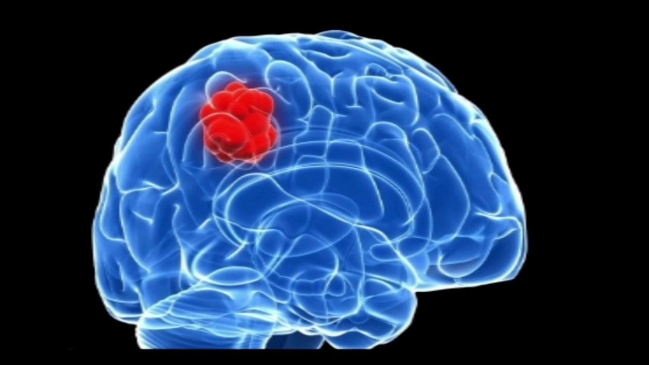
MNPs are the most advanced contrast labels currently being used in research and development for medical imaging. Dr. Shan Wang’s group at Stanford University has developed superparameganetic iron oxide nanoparticles (SPIONs) and fluorescent tag conjugated SPIONs for biological molecular imaging (US 7,682,838 and US 8,722,017 ). Dr. Miqin Zhang’s group at the University of Washington has developed MNPs with a Fe3O4 core and a mesoporous silica shell embedded with carbon dots and paclitaxel (a common anti-cancer drug), and covered by another layer of silica. These MNPs enable confocal and twophoton fluorescence imaging via carbon dots and MRI via magnetic Fe3O4. They also deliver the paclitaxel to cancer cells to kill them through combined photothermal and chemotherapy. Dr. Zhang also developed major histocompatibility complex (MHC) conjugated MNPs for imaging T cells and also chitosan-polyethylene oxide oligomer copolymer coated MNPs for brain tumor imaging and drug delivery (US 20160193369, US 20150320890, and US 20140286872). Dr. Koichiro Hayashi demonstrated the advantages of using SPIONs for cancer theranostics by combining MRI and magnetic hyperthermia treatment (WO/2012/026194). His team modified the SPION clusters with folic acid and polyethylene glycol (PEG) to promote the accumulation of clusters in tumors. Dr. Qun Zhao at the University of Georgia developed hyperthermia treatment of head and neck cancers in a mouse model via intratumor injection of SPIONs. Ultrasmall superparamagnetic iron oxide nanoparticles (USPIONs) having smaller size in diameter, resulting in longer circulation time. These particles can accumulate in the microvascularture before being endocytosed (i.e. removed) by macrophages. Therefore, these particles can be used for tumor-associated microvessel imaging. Dr. Edward Neuwelt reported clinical data with enhanced brain tumor imaging by USPIONs. Other groups from France and Switzerland also reported similar results.
Summary
Magnetic nanoparticles are not only used as MRI contrast labels for medical imaging, but also used as therapeutic drug delivery carriers, as hyperthermia tools, and even as combined drug delivery and imaging agents for cancer therapy. In the next installment, we will discuss further details on the application of these particles in cancer therapeutics.
| Patent Number | Title | Assignee | Inventor |
| US 8,945,628 | Magnetic nanoparticles | The General Hospital Corporation | Ralph Weissleder; Hakho Lee; Tae-Jong Yoon
|
| US 7,462,446 | Magnetic nanoparticle compositions and methods | University of Washington | Miqin Zhang; Nathan Kohler; Jonathan Whitney Gunn
|
| US 6,615,063 | Fluorescence-mediated molecular tomography | The General Hospital Corporation | Vasilis Ntziachristos; Ralph Weissleder
|
| US 8,569,078 | Magnetic-nanoparticle conjugates and methods of use | The General Hospital Corporation | Lee Josephson; Ralph Weissleder; J. Manuel Perez
|
| US 9,097,644 | Magnetic resonance-based viscometers and methods | Massachusetts Institute of Technology,
The General Hospital Corporation |
Lee Josephson; Rui Hong; Michael J. Cima; Ralph Weissleder
|
| US 7,682,838 | Magnetic nanoparticles, magnetic detector arrays, and methods for their use in detecting biological molecules | The Board of Trustees of the Leland Stanford Junior University | Shan X. Wang; Robert L. White; Chris D. Webb; Guanxiong Li
|
| US 8,722,017 | Fluorescent magnetic nanoprobes, methods of making, and methods of use | The Board of Trustees of the Leland Stanford Junior University | Aihua Fu; Shan X. Wang; Sanjiv Sam Gambhir
|
| US 20140286872 | NANOPARTICLE FOR TARGETING BRAIN TUMORS AND DELIVERY OF O6-BENZYLGUANINE | University of Washington | Miqin Zhang; Richard G. Ellenbogen; Forrest Kievit; John R. Silber; Zachary Stephen; Omid Veiseh
|
| US 20150320890 | NANOPARTICLES FOR BRAIN TUMOR IMAGING | University of Washington | Miqin Zhang; Conroy Sun; Omid Veiseh; Narayan Bhattarai
|
| US 20160193369 | MAGNETIC NANOPARTICLE AND METHOD FOR IMAGING T CELLS | University of Washington | Miqin Zhang; Jonathan Whitney Gunn; Cassian Yee
|
| WO/2012/026194 | METAL OXIDE NANOPARTICLE STRUCTURE AND PRODUCTION METHOD FOR SAME | THE UNIVERSITY OF TOKUSHIMA | Koichiro HAYASHI |
– Jing Zhou, PhD and Anthony D. Sabatelli, PhD, JD
This article is for informational purposes, is not intended to constitute legal advice, and may be considered advertising under applicable state laws. The opinions expressed in this article are those of the author only and are not necessarily shared by Dilworth IP, its other attorneys, agents, or staff, or its clients.