Nanomedicine: A Vast Horizon on a Molecular Landscape – Part X, Magnetic Nanoparticles theranostics II
May 16th, 2017 by Jing Zhou | News | Recent News & Articles |
This is the tenth article in a review series on Nanomedicine. We started the series by reviewing the major research areas and entrepreneurial developments in nanomedicine and the relevant patent landscape (Part I and Part II). Following that, we discussed organs-on-a-chip (Part III and Part VIIII), nanotechnology in medical therapeutics: nanoparticles for drug delivery (Part IV), cancer therapeutics (Part V), and bio-imaging (Part VI), and nanoparticles with specific functions: quantum dots for bioimaging and therapy (Part VII) and magnetic nanoparticles for diagnosis (Part VIII). Here, we continue review of the theranostic applications and IP landscape of magnetic nanoparticles (MNPs). As in the past, those patent documents cited in the article are summarized in the table at the end.
MNPs as a dual modality for cancer imaging
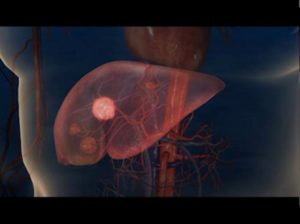
Magnetic nanoparticles are superior imaging contrast agents for Magnetic Resonance Imaging (MRI) due to the intrinsic magnetic properties of nanoparticles. As of 2012, the FDA has approved several MNPs as MRI contrast agents or therapeutic agents: ferumoxides (also known as Feridex in the USA) as an MRI contrast agent for imaging liver lesions; ferucarbotran (also known as Resovist) as MRI contrast agent for imaging liver lesions; ferumoxsil (also known as GastroMARK or Lumirem) as an orally administered MRI contrast agent; and ferumoxytol (also known as Feraheme) as an intravenously administered nanoparticle to treat iron deficiency in adults with chronic kidney disease.
With surface molecular modification, MNPs can be functionalized with suitable fluorescent dyes and radionuclides to enable multimodal imaging, for example, optical imaging, Positron Emission Tomography (PET) imaging and Computed Tomography (CT) imaging. The advantage of multimodal imaging helps to ensure the conformance of cancer diagnosis through the combination of complementary strengths of different imaging techniques. Dr. Gang Bao at Rice University and Dr. Shuming Nie at Emory University developed fluorescent label conjugated magnetic iron oxide nanoparticles for deep-tissue imaging (US 7,459,145). Dr. Anna Moore at Harvard Medical School developed a gold coated iron oxide nanoparticle with a further dextran coating layer for dual modality magnetic resonance imaging (MRI) and surface-enhanced Raman scattering (SERS) imaging (US 8,563,043). Dr. Rafael T.M. de Rosales at King’s College London conjugated a 64Cu radiolable with dithiocarbamate (DTC) and bisphosphonates (BP) to form a [64Cu(dtcbp)2] complex. This complex was further labeled with clinically available dextran-coated superparamagnetic iron oxide nanoparticles (SPIONs) for MRI/PET dual modality imaging (WO2011151631). In vivo studies with these particles in the lymphatic system successfully detected the early spread of cancer. Dr. Weibo Cai at the University of Wisconsin-Milwaukee and Dr. Shaoqin Gong at the University of Wisconsin-Madison developed a water-soluble SPION with 64Cu chelators for MRI/PET dual modality imaging. These nanoparticles were conjugated with cRGD peptides (i.e. a tripeptide of arginine, glycine, and aspartic acid) to target tumors with integrin avb3 expression and to also carry an anticancer drug for targeted tumor treatment.
MNPs for targeted drug delivery
MNPs can accumulate at a target tissue through an enhanced permeability and retention (EPR) effect. Beyond this passive targeting, the surface molecular modification of MNPs enables the active targeting at specific biomarkers of malignant tissues. The multifunctionality of MNPs allows the selective delivery of drugs to the desired location for therapy. Dr. J. Manuel Perez at the University of Central Florida used a co-encapsulation strategy to coat both a near infrared (NIR) dye and a chemotherapeutic agent, taxol, with polyacrylic acid (PAA) on SPIONs. These SPIONs were further conjugated with a folic acid ligand to target folate expressing cancer cells. This combination enabled a theranostic with MRI/optical dual modality imaging and cancer cell targeting (US 8,821,837 and US 8,372,944). Dr. Xiaoyuan Chen at the National Institutes of Health conjugated the anti-cancer drug, doxorubicin (DOX), with a human serum albumin (HAS) coated iron oxide nanoparticle. In a murine breast cancer model, the modified MNPs induced tumor reduction and demonstrated a better therapeutic effect than a DOX only treatment. Dr. Michael Welch and Dr. Wooley Karen at Washington University developed a shell-crosslinked knedel (SCK) nanoparticle with peptide nucleic acids (PNAs) to enhance cell uptake of the nanoparticles and facilitate drug delivery (US 8,354,093).
MNPs for localized hyperthermia treatments
Another unique feature of MNPs is the hyperthermia effect that can be induced under an alternating magnetic field. When the external magnetic field is oscillating, the MNPs continuously rotate to align with the magnetic field. Under this circumstance, the MNPs absorb electromagnetic energy and transform it into heat energy, locally increasing the temperature of their surroundings. Therefore, the temperature of tumor cells targeted by such MNPs can be increased in the range of 43-47 oC and undergo intra- and extracellular degradation mechanisms causing cell death. The advantage of MNP induced hyperthermia is that it is highly localized and has minimal effect on nearby healthy tissues.
Dr. Jinwoo Cheon at Yonsei University synthesized CoFe2O4@MnFe2O4 core-shell nanoparticles and administered these nanoparticles to mice with xenografted human brain cancer cells. The magnetic hyperthermia treatment by these core-shell MNPs demonstrated better results on tumor elimination compared to Feridex. Dr. Cheon’s particle also provided effective hyperthermia treatment versus control groups, showing a similar effect to core-shell MNPs conjugated to doxorubicin (US 8,066,969). Dr. Matthew Basel synthesized paramagnetic iron/iron oxide nanoparticles and loaded these into mouse monocyte/macrophage-like cells to target tumor cells. These MNPs specifically targeted pancreatic tumors and induced localized hyperthermia for cancer treatment (US 20120157824). Dr. James Hainfeld applied MNPs through intravenous injection to target subcutaneous squamous cell carcinoma in mice. An alternating external magnetic field was used to induce hyperthermia to ablate the tumor cell while leaving the surrounding healthy tissue intact (US 7,906,147).
In 2013, MagForce, a German company, announced the approval by the European Medicines Agency (EMA) of a new product, NanoTherm, the treatment of primary or recurrent glioblastoma multiforme, which is a lethal brain tumor with limited treatment options. The new treatment depends on direct injection of the MNPs to tumors and localized hyperthermia for delivering the cancer treatment (US 9,345,768). Clinical trial in 66 patients with recurrent glioblastoma multiforme showed longer overall survival with MNP treatment. Currently NanoTherm has been released in 27 European countries.
Summary
Besides the currently approved MNPs by FDA as MRI contrast agents or therapeutic agents, researchers and scientists are actively developing new MNPs with combined imaging and therapeutic functions to take advantage of the theranostic property of MNPs to enhance clinic outcomes. We are expecting more new products to be clinically approved in the coming years.
| Patent Number | Title | Assignee | Inventor |
| US 7,459,145 | Multifunctional magnetic nanoparticle probes for intracellular molecular imaging and monitoring | Georgia Tech Research Corporation, Emory University |
Gang Bao; Shuming Nie; Nitin Nitin; Leslie LaConte
|
| US 8,563,043 | Innately multimodal nanoparticles | The General Hospital Corporation | Zdravka Medarova; Anna Moore; Mehmet Yigit |
| WO2011151631 | NANOPARTICLES AND THEIR USES IN MOLECULAR IMAGING | KING’S COLLEGE LONDON | Philip, John BLOWER; Mark GREEN; Maite JAUREGHI-OSORO; Rafael TORRES MARTIN DE ROSALES; Peter WILLIAMSON |
| US 8,821,837 | Aqueous method of making magnetic iron oxide nanoparticles | University of Central Florida Research Foundation, Inc. | Jesus Manuel Perez; Sudip Nath |
| US 8,372,944 | Synthesis of hyperbranched amphiphilic polyester and theranostic nanoparticles thereof | University of Central Florida Research Foundation, Inc. | J. Manuel Perez; Santimukul Santra |
| US 8,354,093 | Cell permeable nanoconjugates of shell-crosslinked knedel (SCK) and peptide nucleic acids (“PNAs”) with uniquely expressed or over-expressed mRNA targeting sequences for early diagnosis and therapy of cancer | Washington University | Matthew L. Becker; Huafeng Fang; Xiaoxu Li; Dipanjan Pan; Raffaella Rossin; Xiankai Sun; John-Stephen Taylor; Jeffrey L. Turner; Michael John Welch; Karen L.Wooley |
| US 8,066,969 | Preparation method of magnetic and metal oxide nanoparticles | Industry-Academic Cooperation Foundation, Yonsei University | Jin-Woo Cheon; Jung-Wook Seo; Jae-Hyun Lee |
| US20120157824 | MRI AND OPTICAL ASSAYS FOR PROTEASES | NANOSCALE CORPORATION; KANSAS STATE UNIVERSITY RESEARCH FOUNDATION |
Stefan H. Bossmann; Deryl Troyer; Matthew T. Basel; Thilani Nishanthika Samarakoon; Hongwang Wang; Viktor Chikan; Franklin Orban Kroh; Olga Barbara Koper; Ray Brandon Walker; Xiaoxuan Leaym |
| US 7,906,147 | Functional associative coatings for nanoparticles | Nanoprobes, Inc. | James F. Hainfeld |
| US 9,345,768 | Nanoparticle/active ingredient conjugates | MAGFORCE AG | Andreas Jordan; Norbert Waldoefner; Klaus Decken; Regina Scholz |
– Jing Zhou, PhD and Anthony D. Sabatelli, PhD, JD
This article is for informational purposes, is not intended to constitute legal advice, and may be considered advertising under applicable state laws. The opinions expressed in this article are those of the author only and are not necessarily shared by Dilworth IP, its other attorneys, agents, or staff, or its clients.


